New materials allow light sheet microscopes to see super-resolution

SAN DIEGO, June 4, 2021 — A hyperbolic metamaterial developed at the University of California, San Diego enables ordinary light-sheet microscopes to watch subcellular structures — in other words, enabling a standard microscope to ascertain in superresolution. the fabric, made from nanometers-thin alternating layers of silver and silica glass, shortens the wavelength of sunshine because the sample is illuminated, which allows the microscope to ascertain beyond the diffraction limit.
In the application, the light-shrinking material converts low-resolution light to high-resolution light, said Zhaowei Liu, a professor of electrical and computer engineering at UC San Diego. “It’s very simple and straightforward to use. Just place a sample on the fabric, then put the entire thing under a traditional microscope — no fancy modification needed," Liu said.
The work overcomes a serious limitation of conventional light microscopes, also as a limitation in superresolution imaging methods more generally. Light microscopes are useful for the observation of live cells, and other methods, like microscopy, allow the observer to ascertain subcellular structures, though the sample must be placed during a chamber. Other methods involve altering the cells, like expansion microscopy. Light microscopy allows researchers to look at living cells in their wild, though conventional light microscopes, with a resolution of 200 nm, are unable to watch objects closer than that distance as separate objects.
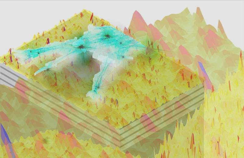
The technology developed by the University of California team combines these two functions. With the hyperbolic metamaterial in situ, coated on a slide, a standard microscope is in a position to ascertain subcellular structures with a resolution of up to 40 nm. As light passes through, its wavelengths shorten and scatter to get a series of random high-resolution speckled patterns. When a sample is mounted on the slide, it gets illuminated in several ways by this series of speckled light patterns, creating a series of low-resolution images. the pictures are captured then pieced together by a reconstruction algorithm to supply a high-resolution image.
The researchers tested the technology with a billboard inverted microscope. They were ready to image fine features, like actin filaments, in fluorescently labeled Cos-7 cells, which aren't discernable when using the microscope alone. The technology also enabled the researchers to obviously distinguish tiny fluorescent beads and quantum dots that were spaced 40 to 80 nm apart.

